Neuroimaging insights into the brain networks of induced migraine attacks
Neuroimaging studies have changed the way we understand migraine and cluster headache, supporting a key role of the brain in their pathophysiology.1 Functional magnetic resonance imaging (fMRI) is a functional neuroimaging procedure based on MRI that is able to measure variations in brain activity by detecting local changes in blood flow.2 At the recent virtual 6th Congress of the European Academy of Neurology (EAN) held 23–26 May 2020, Dr. Daniele Martinelli (Mondino National Neurological Institute Foundation in Pavia, Italy) spoke in a session on “Headache and Pain.” In his presentation entitled “Brain networks in migraine: A pilot study using advanced fMRI techniques in experimentally-induced attacks,” Dr. Martinelli discussed the latest pilot study results in applying advanced fMRI technique to evaluate the pain process during provoked migraine attacks.
A pain model that captures pattern fMRI activity within and across brain regions can move us forward in understanding the neurological basis of pain and hopefully in finding a reliable biomarker for migraine.
There is a high complexity of network interplay during migraine
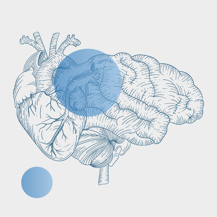
Dr. Martinelli expressed that current research has identified the different structures involved in migraine, including pain modulation in the brainstem and the processing of sensory input by the thalamus and the neocortex. However, the interplay between these structures is not clearly understood.
Since the seminal positron emission tomography (PET) study by Weiller et al. in 1995,3 several MRI studies have described the main brain regions involved in the various migraine phases.1 Dr. Martinelli articulated that there is complexity in the various brain regions involved during a migraine attack; however, these known brain activities only capture a single phase of a migraine attack and there is a gap in understanding the comprehensive evaluation of this complex event.
A pilot study to evaluate brain activity in migraine
Dr. Martinelli explained that the aim of their study was to evaluate the brain activity in each phase of an induced headache attack in episodic migraineurs with the use of advanced fMRI techniques. As is well-described in literature,4 the nitroglycerin paradigm was used where an oral nitroglycerin administration causes a migraine attack and pain is only quenched with an anti-inflammatory drug once patients reach a pain intensity of 5 out of 10.
According to Dr. Martinelli, 10 patients were enrolled, 5 with episodic migraine without aura that experience drug-induced migraine attack with clinical characteristics and 4 healthy subjects as controls. Data were analyzed with seed based component analysis. The mean effect for each phase was normalized to its baseline and a functional connectivity quantification was performed to rank the strongest difference in coupling between brain regions during a migraine attack.
Functional connectivity among brain regions during migraine
Based on the results of this study, Dr. Martinelli concluded that the brainstem elements involved in the pain circuitry and the thalamus depended on the migraine cycle and exhibited an outer functional coupling, particularly during the prodromal phase. Dr. Martinelli further emphasized that the thalamus strongly altered its coupling with the frontal, temporal, and cerebral cortex and therefore showed a greater involvement during the full-blown phase. Therefore, Dr. Martinelli stressed that there is a key role in the interaction between the brainstem circuitry and the thalamus-hypothalamus axis, which suggests that a migraine attack is not led by a single brainstem generator, but rather a more complex process as a cyclical oscillation between networks.
fMRI in research identifies brain regions to enable better migraine care
Dr. Martinelli summarized that using fMRI and a pain model to capture brain activity could help elucidate the neurological basis of pain. According to Dr. Martinelli, establishing a variety of techniques and approaches will clarify migraine pain signature and help find a reliable biomarker. These approaches will help address the needs of patients, predict their progression and their response to a particular treatment.
Messina R, Filippi M, Goadsby PJ. Recent advances in headache neuroimaging: Curr Opin Neurol. 2018;31(4):379–85.
Lovati C, Giani L, Mele F, et al. Brain plasticity and migraine transformation: fMRI evidences. Expert Rev Neurother. 2016;16(12):1413–25.
Weiller C, May A, Limmroth V, et al. Brain Stem Activation in Spontaneous Human Migraine Attacks. Nat Med. 1995;1(7):658–660.
Demartini C, Greco R, Zanaboni AM, et al. Nitroglycerin as a comparative experimental model of migraine pain: From animal to human and back. Prog Neurobiol. 2019;177:15–32.